

Patients often ask me whether I perform the nerve-burning procedure.
My usual answer is something along the lines of:
Possibly—but first we need to decide which nerve is involved, why we believe it is causing your pain, and whether we should heat it, pulse it, cool the electrode, treat it inside the bone, or leave it completely alone.
Radiofrequency ablation, commonly called RFA, is not one single procedure. It is a family of treatments that use radiofrequency energy in different ways to interrupt or modify pain signals.
Calling every radiofrequency procedure nerve burning is a little like calling every surgery cutting something. There may be a small amount of technical truth in the description, but it leaves out almost everything that determines whether the treatment is appropriate.
Conventional thermal RFA, cooled RFA, pulsed radiofrequency and basivertebral nerve ablation involve different targets, different temperatures, different biological effects and very different levels of scientific evidence.
The most important question is therefore not:
Does RFA work?
The better questions are:
- What structure is actually generating the pain?
- Which nerve carries the pain signal from that structure?
- How confident are we in the diagnosis?
- Which type of radiofrequency treatment fits that nerve?
- What does the evidence show for that specific condition?
That distinction matters because RFA can be extremely effective when the correct patient, diagnosis and anatomical target come together. It is much less impressive when it is used simply because an MRI shows arthritis or because pain has persisted for a long time.
What Is Radiofrequency Ablation?
Radiofrequency procedures use an insulated needle, called a cannula, placed near a specific nerve or within a specific anatomical target under image guidance.
An electrode is inserted through the cannula. Radiofrequency energy is then delivered through the electrode.
Depending on the technique, that energy may:
- Create a controlled thermal lesion in a small sensory nerve
- Produce a larger treatment zone around an anatomically variable nerve
- Apply electrical fields in pulses without intentionally destroying the nerve
- Treat a nerve located inside a vertebral body
Most procedures are performed using fluoroscopy, or real-time X-ray guidance. Ultrasound or CT guidance may be used for certain peripheral nerve targets.
The goal is not to remove normal sensation from a large area or damage a major nerve. The goal is to precisely treat a small nerve that has been identified as carrying a chronic pain signal.
The Main Types of Radiofrequency Treatment
The major categories include:
- Conventional thermal radiofrequency ablation
- Cooled radiofrequency ablation
- Pulsed radiofrequency
- Basivertebral nerve ablation
- Bipolar or multipolar radiofrequency techniques
These approaches are related, but they should not be treated as interchangeable.
1. Conventional Thermal RFA
Conventional RFA uses continuous radiofrequency energy to create a small, controlled area of heat around the active portion of the electrode.
This is the technique most commonly used for medial branch nerves supplying painful facet joints in the neck or lower back.
Where Conventional RFA May Be Used
Common targets include:
- Cervical medial branch nerves for facet-mediated neck pain
- The third occipital nerve for selected cervicogenic headaches
- Thoracic medial branch nerves in carefully selected patients
- Lumbar medial branch nerves for facet-mediated low-back pain
- Selected peripheral sensory nerves
Facet Joint Pain
Facet joints are small joints located along the back of the spine. They provide stability and help control movement.
Like other joints, they may become arthritic, inflamed or painful. However, facet arthritis on an MRI does not prove that the facet joint is causing a patient's pain.
This is an important point because imaging abnormalities are common, particularly as we get older. An MRI can show us what a joint looks like, but it cannot tell us with certainty whether that joint is producing pain.
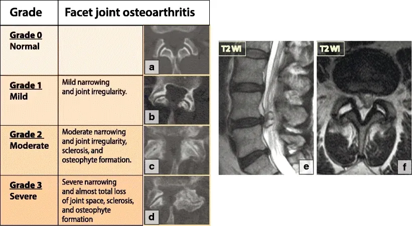
Facet arthritis can range from mild to severe on imaging, but the grade alone does not prove that the joint is the source of pain. (Image: Irina Nefedova, CC BY 4.0)
Why Diagnostic Medial Branch Blocks Matter
Before performing a medial branch RFA, physicians commonly perform one or more diagnostic medial branch blocks.
During a medial branch block, a small amount of local anesthetic is placed around the nerves carrying sensation from the suspected facet joints. The patient then tracks how much relief occurs and how long it lasts.
A clearly positive response increases confidence that the facet joints are meaningful pain generators and that RFA may help.
The diagnostic block is not a perfect test. False-positive and false-negative results can occur. There is also continuing debate about whether one or two blocks should be required and exactly what percentage of relief should qualify as a positive response.
Still, diagnostic blocks are generally more useful than choosing patients based on MRI findings alone.
In many cases, the actual ablation is the straightforward part. Correctly identifying the nerve worth treating is where most of the medicine happens.
What Does the Evidence Show for Facet RFA?
Facet-mediated spinal pain has one of the most established evidence bases for conventional RFA.
A 2015 Cochrane review found moderate-quality evidence that facet radiofrequency denervation reduced pain more than placebo in the short term. It also found evidence of greater effectiveness than steroid injections at several follow-up periods.[1]
However, the literature is not uniformly positive.
A major 2026 systematic review and meta-analysis evaluated 55 randomized trials involving 4,394 patients with facet, sacroiliac, discogenic or nonspecific chronic low-back pain. Radiofrequency denervation produced statistically significant but generally small average improvements in pain and function. The average differences did not exceed the authors' predefined thresholds for minimum clinically important differences.[2]
That deserves an honest explanation.
It does not necessarily mean that no individual patient benefits meaningfully. Clinical trials report average results across groups, and those groups may include different selection protocols, nerve targets, techniques, lesion sizes and definitions of success.
It does mean that RFA should not be marketed as a universal solution for chronic back pain.
The evidence is most applicable when:
- The pain pattern is compatible with facet-mediated pain
- Other major pain generators have been considered
- Diagnostic blocks produce convincing temporary relief
- The procedure is performed using appropriate anatomical and technical standards
2. Cooled Radiofrequency Ablation
Cooled RFA is another thermal technique, despite its somewhat confusing name.
The tissue is still heated. The difference is that water circulates through the electrode and cools the tip internally. This prevents the immediate tissue touching the electrode from overheating too quickly.
As a result, radiofrequency energy can spread farther into the surrounding tissue and create a larger, more spherical lesion.
Think of it less as making the procedure cold and more as controlling the temperature at the needle tip so a larger treatment zone can develop.
Why Would a Larger Lesion Be Helpful?
Some nerves travel along relatively predictable anatomical pathways. Others have more variable courses or divide into multiple small branches.
A larger lesion may increase the probability of capturing the intended sensory nerve branches when the anatomy is variable.
However, larger does not automatically mean better. A larger treatment zone must still be carefully positioned to avoid motor nerves, blood vessels and other structures.
Where Cooled RFA May Be Used
Common applications include:
- Genicular nerves around the knee
- Sacral lateral branches involved in posterior SI joint pain
- Other anatomically variable sensory nerve targets
Cooled RFA for Sacroiliac Joint Pain
The sacroiliac joints connect the sacrum to the pelvis. SI joint pain may cause low-back, upper-buttock, groin or posterior thigh discomfort.
The posterior portion of the SI joint complex receives sensory innervation through branches that may include the L5 dorsal ramus and sacral lateral branches.
These nerves can be anatomically variable, which makes lesion size and technique especially important.
What Does the Evidence Show?
A multicenter randomized comparative-effectiveness trial involving 210 patients compared cooled RFA with standard medical management. At three months, the cooled-RFA group had a mean pain-score reduction of approximately 2.5 points, compared with about 0.4 points in the standard-treatment group. Functional and quality-of-life outcomes also favored cooled RFA.[3]
Follow-up from the study showed sustained clinical utility at 12 months, with approximately 57% of treated patients meeting the study's responder criteria.[4]
A 2026 network meta-analysis of 18 randomized trials involving 1,075 participants found that radiofrequency-based interventions generally ranked ahead of steroid injections, sham procedures and conservative treatment for SI joint pain. However, the certainty of evidence was often rated low or very low because of differences among studies and limited direct comparisons.[5]
Current pain-society guidance supports sacral lateral branch RFA for properly selected patients, while also recognizing that diagnostic protocols, electrode placement and lesion techniques vary.[6,7]
Who May Be Considered?
A reasonable candidate may have:
- Pain in a pattern compatible with SI joint complex pain
- Positive findings during a careful physical examination
- Failure of appropriate conservative treatment
- Meaningful temporary relief from image-guided diagnostic SI joint or lateral branch blocks
- No better explanation for the symptoms
No individual examination maneuver or MRI finding definitively diagnoses SI joint pain. The diagnosis is usually built from several pieces of evidence.
3. Basivertebral Nerve Ablation
Basivertebral nerve ablation, or BVNA, is substantially different from conventional facet RFA.
The basivertebral nerve is located inside the vertebral body. It carries pain signals arising from damaged vertebral endplates.
The vertebral endplates sit between the spinal discs and the vertebral bodies. These endplates can become damaged and inflamed as part of the degenerative process.
This produces a specific condition known as vertebrogenic low-back pain.
What Is Vertebrogenic Pain?
Vertebrogenic pain is generally characterized by chronic, primarily axial low-back pain associated with endplate damage.
Patients may describe:
- Deep pain near the center of the lower back
- Pain made worse by sitting, bending or loading the spine
- Pain when moving from sitting to standing
- Symptoms that are primarily in the back rather than the leg
- Limited improvement with appropriate conservative treatment
MRI commonly shows Type 1 or Type 2 Modic changes at the vertebral endplates.
Type 1 Modic changes generally reflect inflammation and edema within the vertebral bone marrow. Type 2 changes reflect fatty replacement associated with chronic endplate degeneration.
Modic changes do not automatically prove that someone has vertebrogenic pain. The symptoms and MRI findings must fit together.
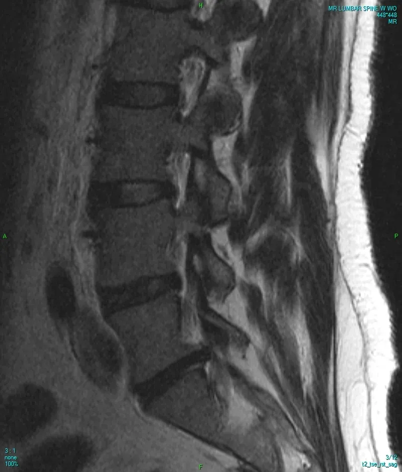
MRI is one part of a vertebrogenic-pain evaluation. Endplate findings must still match the patient's symptoms and examination.
How Is BVN Ablation Performed?
A small access channel is created through the pedicle into the vertebral body. A radiofrequency probe is then positioned near the basivertebral nerve inside the bone.
The nerve is treated with thermal energy to interrupt pain signaling from the damaged endplates.
The commonly studied treatment levels are L3 through S1.
Unlike a medial branch RFA, this is an intraosseous procedure. It does not target the facet joints, the spinal nerve roots or the disc itself.
What Does the Evidence Show?
The evidence for basivertebral nerve ablation has expanded rapidly.
In the randomized INTRACEPT trial, BVN ablation was compared with standard nonsurgical care in carefully selected patients with chronic vertebrogenic pain and Modic Type 1 or Type 2 changes.
At 12 months, patients treated with BVN ablation had an average Oswestry Disability Index improvement of 25.7 points and an average pain reduction of 3.8 centimeters on a 10-centimeter visual analog scale. Sixty-four percent achieved at least a 50% reduction in pain, and 29% reported being pain-free.[8]
A separate randomized, sham-controlled trial also demonstrated significantly greater functional improvement after BVN ablation than after a sham procedure.[9]
Long-term durability is particularly important because many pain procedures are studied for only a few months.
A pooled five-year analysis included 249 patients from three prospective clinical trials. At five years, average pain scores had improved by 4.32 points and disability scores by 28 points compared with baseline. Approximately 32% reported being pain-free.
Among patients taking opioids at baseline, 65.2% were no longer taking them at five years. The number of spinal injections also decreased substantially.[10]
A prospective real-world cohort published in 2026 found that benefits extended beyond the original tightly controlled trials. At 12 months, approximately 53% met the study's disability-response threshold and approximately 68% achieved at least a two-point improvement in pain.[11]
Professional guidance from the American Society of Pain and Neuroscience supports BVN ablation for properly selected patients with vertebrogenic pain.[12]
An economic analysis also estimated an incremental cost-effectiveness ratio of approximately $11,376 per quality-adjusted life-year gained compared with standard care, which is well below commonly used United States cost-effectiveness thresholds.[13]
The Important Caveat
The BVN literature is encouraging, but transparency matters.
The major clinical trials have had industry sponsorship or manufacturer involvement. That does not invalidate the findings, particularly when multiple prospective trials show durable and consistent outcomes. However, independent confirmatory trials would strengthen confidence and help clarify how results translate across broader clinical settings.
BVN ablation should not be offered merely because an MRI report contains the word Modic.
The best candidate has:
- Chronic axial low-back pain, generally lasting at least six months
- Failure of appropriate nonsurgical care
- A clinical pattern compatible with vertebral endplate pain
- Type 1 or Type 2 Modic changes at appropriate lumbar levels
- No competing diagnosis that better explains the symptoms
4. Pulsed Radiofrequency
Pulsed radiofrequency, or PRF, is different from conventional thermal ablation.
Instead of continuously applying energy to create a controlled destructive lesion, PRF delivers brief bursts of high-voltage electrical current separated by inactive cooling periods.
The temperature near the electrode is typically limited to approximately 42°C.
The purpose is primarily neuromodulation rather than intentional nerve destruction.
In other words, conventional RFA attempts to interrupt a pain pathway by creating a thermal lesion. PRF attempts to alter the way the nerve processes or transmits pain without creating the same degree of structural injury.
Why Use Pulsed RF?
PRF may be attractive when the target is a mixed nerve, a dorsal root ganglion or another structure where destructive thermal treatment could produce weakness, numbness or other unwanted deficits.
Potential applications include:
- Cervical and lumbar radicular pain
- Trigeminal neuralgia
- Postherpetic neuralgia
- Occipital neuralgia
- Suprascapular nerve pain
- Intercostal neuralgia
- Cluneal neuralgia
- Meralgia paresthetica
- Pudendal neuralgia
- Coccygodynia
- Selected headache disorders
- Other focal neuropathic pain syndromes
How Might PRF Work?
The exact mechanism is still being studied.
PRF appears to affect electrical signaling, gene expression, synaptic activity and inflammatory pathways within pain-processing neurons.
Recent preclinical research suggests PRF may inhibit axonal transport within smaller dorsal-root-ganglion neurons and decrease the release of pain-related neuropeptides, including calcitonin gene-related peptide and substance P.[14,15]
This does not mean the nerve is simply reset, a phrase often used casually. The biological effects are complex and likely differ depending on the nerve, voltage, pulse duration and treatment time.
Pulsed RF for Radicular Pain
PRF applied near the dorsal root ganglion has been studied for both cervical and lumbar radicular pain.
The dorsal root ganglion contains sensory nerve-cell bodies and plays an important role in neuropathic and radicular pain signaling.
Multiple randomized trials and reviews suggest PRF can reduce radicular pain in selected patients, often without the risks associated with creating a destructive lesion near a spinal nerve.[16,17]
However, studies use different voltages, treatment durations and outcome measures. PRF should therefore be described as promising rather than universally proven.
Evidence Assessment
The evidence for PRF in radicular pain is meaningful and growing, but it is less standardized and less consistent than the evidence for medial branch RFA in properly diagnosed facet pain or BVN ablation in carefully selected vertebrogenic pain.
Pulsed RF for Trigeminal Neuralgia
Trigeminal neuralgia causes severe, electric shock-like facial pain.
Because the trigeminal nerve carries important facial sensation, nondestructive or minimally destructive strategies are attractive.
A multicenter, randomized, double-blind trial involving 134 patients found that high-voltage PRF produced a one-year response rate of 73.1%, compared with 32.8% after nerve block treatment.[18]
A retrospective series of 149 patients reported a median recurrence-free survival of 118 months, although retrospective findings should be interpreted more cautiously than randomized data.[19]
A 2025 randomized trial found that combining PRF with low-temperature continuous RFA improved 12-month response rates compared with PRF alone.[20]
These results are encouraging, although treatment protocols and patient populations may differ considerably among studies.
Pulsed RF for Postherpetic Neuralgia
Postherpetic neuralgia is persistent neuropathic pain following shingles.
Reviews consistently identify this as one of the better-supported PRF applications. PRF may be applied near the affected dorsal root ganglion or peripheral nerve, depending on the pain distribution and clinical situation.[16]
Pulsed RF for Occipital Neuralgia
Occipital neuralgia causes pain arising from the occipital nerves at the back of the head.
PRF has been studied as a treatment when diagnostic nerve blocks provide temporary relief. Clinical evidence suggests PRF may provide longer benefit than steroid injection alone in selected patients.[17]
Pulsed and Cooled RF for Knee Osteoarthritis
Genicular nerves are small sensory nerves that carry pain signals from the knee joint.
Genicular nerve RFA does not repair cartilage, reverse arthritis or mechanically stabilize the knee. Its purpose is to decrease pain sufficiently to improve walking, sleep, exercise and daily function.
It may be considered for:
- Symptomatic knee osteoarthritis
- Patients delaying knee replacement
- Patients who are not surgical candidates
- Persistent pain after knee replacement once infection, loosening, instability and other mechanical problems have been excluded
A 2024 systematic review and meta-analysis included 11 studies and 604 patients. Both cooled and pulsed genicular RF produced significant pain reduction through six months. Pulsed RF also showed functional improvement at one and three months.
However, the review identified a limited number of randomized trials and incomplete functional-outcome reporting.[21]
Therefore, genicular RFA appears useful for selected patients, but technique, target selection and expectations matter.
Pulsed RF for Shoulder Pain
The suprascapular nerve supplies sensation to much of the shoulder joint and also contributes to motor function.
Because it is not purely a sensory nerve, destructive treatment requires caution.
PRF of the suprascapular nerve has shown improvements in pain and shoulder mobility in selected patients with chronic shoulder pain, although the evidence base remains smaller than that for established spinal applications.[16,17]
Other Peripheral Neuralgias
PRF and RFA have also been reported for:
- Cluneal neuralgia
- Intercostal neuralgia
- Meralgia paresthetica
- Pudendal neuralgia
- Neuroma pain
- Postoperative nerve pain
- Coccygodynia
- Selected cancer-related pain
- Other focal peripheral nerve syndromes
A retrospective study of RF treatments for peripheral neuralgias found that approximately 80% of patients reported some improvement. Average pain scores decreased from 6.3 to 3.6, and the average duration of improvement was approximately 129 days.[22]
These results are promising, but they should not be interpreted as equal to high-quality randomized evidence. Peripheral neuralgias are heterogeneous, and many published studies remain small or retrospective.
5. Bipolar and Multipolar RFA
Conventional monopolar RFA uses a single active electrode and a grounding pad.
Bipolar RFA passes current between two electrodes. Multipolar techniques use several electrodes or lesion sites to create a broader or more continuous treatment zone.
Potential applications include:
- SI joint lateral branches
- Genicular nerves
- Other branching peripheral sensory networks
- Anatomical targets where a strip or connected lesion may be useful
These techniques may improve nerve capture in certain anatomical settings, but outcomes depend heavily on electrode spacing, orientation, lesion temperature and nerve anatomy.
Once again, the goal should not be to create the largest possible lesion. The goal is to create an adequately sized lesion in the correct location.
Where Can Radiofrequency Procedures Be Performed?
More Established Applications
The more established RF targets include:
- Cervical medial branch nerves
- Third occipital nerve
- Lumbar medial branch nerves
- Basivertebral nerve
- Genicular nerves
- Sacral lateral branches
Selected or Emerging Applications
Applications with growing but less consistent evidence include:
- Dorsal root ganglion
- Trigeminal nerve
- Occipital nerves
- Suprascapular nerve
- Hip articular branches
- Cluneal nerves
- Intercostal nerves
- Pudendal nerve
- Other focal peripheral neuralgias
The evidence should not be presented as equally strong across every anatomical target.
A procedure can be biologically plausible and clinically helpful without having the same depth of evidence as a treatment studied in several large randomized trials.
How Strong Is the Evidence Overall?
The fairest summary is that the effectiveness of radiofrequency treatment depends heavily on the diagnosis and target.
Stronger or More Established Evidence
- Medial branch RFA for carefully selected facet-mediated pain
- BVN ablation for chronic vertebrogenic pain with compatible Modic changes
- Sacral lateral branch RFA for selected SI joint complex pain
Moderate and Growing Evidence
- Genicular RFA for knee osteoarthritis
- PRF of the dorsal root ganglion for radicular pain
- PRF for postherpetic neuralgia
- PRF or RF strategies for trigeminal neuralgia
- PRF of the occipital or suprascapular nerves
Limited or Emerging Evidence
- Many uncommon peripheral neuralgias
- Discogenic pain treated with conventional RFA
- Nonspecific chronic low-back pain without a clearly identified generator
- Novel nerve targets supported mainly by small observational studies
What the 2026 Meta-Analysis Really Means
The 2026 systematic review deserves special attention because its conclusion may initially seem at odds with more favorable individual trials.
The authors found that RF denervation produced small improvements across several categories of chronic low-back pain, but average between-group differences did not reach predefined clinically meaningful thresholds.[2]
That finding should not be dismissed.
It tells us several important things:
First, radiofrequency treatment should not be applied broadly to everyone with chronic low-back pain.
Second, the term RFA may be too broad to evaluate as though it represents one uniform intervention.
Third, patient selection and procedural technique are likely major determinants of outcome.
A study combining facet pain, SI joint pain, discogenic pain and nonspecific low-back pain is somewhat like evaluating whether medication works without separating antibiotics, blood-pressure medication and insulin.
They are all medications, but the diagnosis determines whether any particular one makes sense.
Similarly, a medial branch RFA, a sacral lateral branch RFA, a pulsed DRG procedure and a basivertebral nerve ablation should not be assumed to have the same effectiveness simply because they all use radiofrequency energy.
Why Patient Selection Matters More Than the Machine
Radiofrequency generators are sophisticated tools, but the machine does not diagnose the patient.
Successful treatment begins with identifying the pain generator.
Depending on the condition, this may require:
- A detailed pain history
- A focused neurological and musculoskeletal examination
- Review of MRI or other imaging
- Diagnostic medial branch blocks
- SI joint injections or lateral branch blocks
- Genicular nerve blocks
- Peripheral nerve blocks
- Correlation of Modic changes with a vertebrogenic pain pattern
- Exclusion of infection, fracture, instability, tumor or progressive neurological disease

Patient selection begins with the history and physical examination, then uses imaging and diagnostic blocks to test the suspected pain source.
A technically perfect ablation of the wrong nerve is still the wrong procedure.
How Long Does RFA Last?
Duration varies by diagnosis, technique and individual patient.
Conventional medial branch nerves may regenerate over time. When that happens, pain may return.
Many patients who respond to medial branch RFA experience relief for approximately six to 18 months, although results may be shorter or longer.
Relief after SI joint or genicular RFA also varies. Some patients obtain months of benefit, while others experience limited improvement.
BVN-ablation studies suggest that benefit can persist for at least five years in appropriately selected patients. This may be partly because the target and disease process differ from those involved in peripheral medial branch neurotomy.
PRF outcomes are especially variable because PRF is neuromodulatory rather than a predictable thermal neurotomy.
Do the Nerves Grow Back?
Some small peripheral sensory nerves treated with conventional RFA can regenerate.
This is one reason the effect is not always permanent.
Nerve regeneration does not necessarily mean the procedure failed. A treatment that provides a meaningful period of improved function, better sleep or reduced medication use may still be valuable.
Repeat RFA may be considered when:
- The original procedure provided substantial and durable relief
- The same pain pattern returns
- The examination remains consistent
- No new diagnosis better explains the symptoms
A repeat diagnostic block may or may not be required depending on the clinical circumstances and insurance rules.
Is RFA Painful?
The skin and deeper tissues are numbed with local anesthetic.
Patients may feel pressure, muscle twitching, temporary stimulation or brief discomfort during needle positioning and testing.
Sedation may be appropriate for selected procedures or patients, but excessive sedation during diagnostic blocks can make the results harder to interpret.
Temporary post-procedure soreness, muscle spasm or neuritis can occur. Relief may not be immediate and sometimes develops over several weeks.
What Are the Risks?
Risks vary by anatomical target and technique but may include:
- Bleeding
- Infection
- Temporary increase in pain
- Local numbness
- Neuritis or burning discomfort
- Allergic reaction
- Injury to a nearby nerve
- Weakness
- Vascular injury
- Skin injury
- Failure to improve pain
Serious neurological complications are uncommon when procedures are performed with appropriate imaging, anatomical knowledge and safety testing.
Patients with pacemakers, spinal cord stimulators or other implanted electrical devices require additional planning. Anticoagulant and antiplatelet medications must also be reviewed individually.
Who Should Not Receive RFA?
RFA may not be appropriate when:
- The pain generator has not been adequately identified
- Symptoms are primarily caused by untreated spinal instability
- There is active infection
- A progressive neurological deficit requires different evaluation
- Pain is caused by a fracture, tumor or other urgent condition
- Diagnostic blocks do not support the proposed target
- The expected benefit does not outweigh the risks
- The procedure is being offered solely because an MRI shows degeneration
Degeneration is common. Pain is personal. The two overlap, but they are not identical.
The Bottom Line
Radiofrequency treatment is not a single procedure, and it should not be judged as though it is.
Conventional thermal RFA works best when a small sensory nerve has been convincingly linked to a painful structure, particularly the spinal facet joints.
Cooled RFA creates a larger treatment zone and may be useful for variable nerve networks such as the sacral lateral branches or genicular nerves.
Pulsed radiofrequency attempts to modify nerve signaling without creating the same thermal lesion, making it useful for selected dorsal root ganglion and peripheral nerve conditions.
Basivertebral nerve ablation is an intraosseous treatment for a specific form of chronic vertebrogenic low-back pain associated with vertebral endplate damage and Modic changes.
At Horizon Spine & Pain of Utah, we do not begin by asking which nerve we can ablate.
We begin by identifying the most likely pain generator.
Only then can we determine whether conventional RFA, cooled RFA, pulsed radiofrequency, basivertebral nerve ablation—or no ablation at all—offers the most reasonable path forward.
Radiofrequency is a toolbox.
The science is not simply knowing how to use the tool. It is knowing when the problem is actually a nail.
Frequently Asked Questions
Is radiofrequency ablation the same as nerve burning?
Conventional RFA does create a controlled thermal lesion, but nerve burning is an oversimplification. Cooled RFA modifies lesion size, PRF is primarily neuromodulatory, and BVN ablation treats a nerve inside the vertebral body.
Which type of RFA is best?
There is no universally best type. The correct technique depends on the diagnosis, nerve anatomy and evidence for that particular condition.
Does RFA fix arthritis?
No. RFA does not reverse arthritis or rebuild cartilage. It reduces pain signaling from selected sensory nerves.
Can RFA help sciatica?
Conventional destructive RFA is generally not performed on a major spinal nerve producing sciatica. Pulsed RF near the dorsal root ganglion may be considered in selected cases because it aims to modify signaling without intentionally destroying the nerve.
Can RFA prevent back surgery?
Sometimes an effective nonsurgical treatment may help a patient delay or avoid surgery. However, RFA is not a substitute for surgery when there is instability, progressive weakness, severe structural compression or another clear surgical indication.
Does insurance cover RFA?
Coverage depends on the diagnosis, anatomical target, prior conservative treatment, diagnostic-block results and the patient's insurance policy. Some applications are widely covered, while newer or less-established applications may not be.
How do I know whether I am a candidate?
A candidacy evaluation should include a careful history, examination, imaging review and, when appropriate, diagnostic blocks. The decision should not be based on an MRI report alone.
References
- [1] Maas ET, Ostelo RW, Niemisto L, et al. Radiofrequency denervation for chronic low back pain. Cochrane Database Syst Rev. 2015;(10):CD008572. doi:10.1002/14651858.CD008572.pub2.
- [2] Truong KMTL, Maas E, Rasmussen MM, et al. Current evidence on radiofrequency denervation for chronic low back pain: a systematic review and meta-analysis. BMJ Open. 2026;16(7):e105106. doi:10.1136/bmjopen-2025-105106.
- [3] Cohen SP, Kapural L, Kohan L, et al. Cooled radiofrequency ablation versus standard medical management for chronic sacroiliac joint pain: a multicenter, randomized comparative effectiveness study. Reg Anesth Pain Med. 2024;49(3):184-191. doi:10.1136/rapm-2023-104568.
- [4] Cohen SP, Kapural L, Kohan L, et al. Cooled radiofrequency ablation provides extended clinical utility in the management of chronic sacroiliac joint pain: 12-month follow-up results. Reg Anesth Pain Med. 2026;51(6):660-669. doi:10.1136/rapm-2024-106315.
- [5] Park JH, Jang JN, Park S, et al. Interventional treatments for sacroiliac joint pain: a systematic review and network meta-analysis. Reg Anesth Pain Med. Published online March 30, 2026. doi:10.1136/rapm-2026-107633.
- [6] Sayed D, Deer TR, Tieppo Francio V, et al. American Society of Pain and Neuroscience best practice guideline for the treatment of sacroiliac disorders. J Pain Res. 2024;17:1601-1638. doi:10.2147/JPR.S464393.
- [7] McCormick ZL, Hurley RW, Anitescu M, et al. Consensus practice guidelines on sacroiliac joint complex pain from a multispecialty, international working group. Pain Med. 2025;26(12):817-917. doi:10.1093/pm/pnaf129.
- [8] Smuck M, Khalil J, Barrette K, et al. Prospective, randomized, multicenter study of intraosseous basivertebral nerve ablation for the treatment of chronic low back pain: 12-month results. Reg Anesth Pain Med. 2021;46(8):683-693. doi:10.1136/rapm-2020-102259.
- [9] Fischgrund JS, Rhyne A, Franke J, et al. Intraosseous basivertebral nerve ablation for the treatment of chronic low back pain: a prospective randomized double-blind sham-controlled multicenter study. Eur Spine J. 2018;27(5):1146-1156. doi:10.1007/s00586-018-5496-1.
- [10] Khalil JG, Truumees E, Macadaeg K, et al. Intraosseous basivertebral nerve ablation: a 5-year pooled analysis from three prospective clinical trials. Interv Pain Med. 2024;3(4):100529. doi:10.1016/j.inpm.2024.100529.
- [11] McCormick ZL, Fogarty AE, Conger A, et al. The effectiveness of basivertebral nerve radiofrequency ablation for the treatment of vertebrogenic low back pain: 1-year results of a prospective real-world cohort study. Pain Med. 2026;27(3):254-261. doi:10.1093/pm/pnaf122.
- [12] Sayed D, Naidu RK, Patel KV, et al. Best practice guidelines on the diagnosis and treatment of vertebrogenic pain with basivertebral nerve ablation from the American Society of Pain and Neuroscience. J Pain Res. 2022;15:2801-2819. doi:10.2147/JPR.S378544.
- [13] Smuck M, McCormick ZL, Gilligan C, et al. A cost-effectiveness analysis of intraosseous basivertebral nerve ablation for the treatment of chronic low back pain. Spine J. 2025;25(2):201-210. doi:10.1016/j.spinee.2024.09.016.
- [14] Yuba T, Koyama Y, Uematsu H, et al. Elucidation of the treatment mechanism of pulsed radiofrequency based on its anti-inflammatory effects. Sci Rep. 2025;15(1):33611. doi:10.1038/s41598-025-19045-z.
- [15] Sam J, Catapano M, Sahni S, et al. Pulsed radiofrequency in interventional pain management: cellular and molecular mechanisms of action—an update and review. Pain Physician. 2021;24(8):525-532.
- [16] Jitsinthunun T, Li C, Ng TK, Zinboonyahgoon N. Pulsed radiofrequency treatment: evidence for and applications in chronic pain. Pain Physician. 2025;28(6):467-481.
- [17] Vanneste T, Van Lantschoot A, Van Boxem K, Van Zundert J. Pulsed radiofrequency in chronic pain. Curr Opin Anaesthesiol. 2017;30(5):577-582. doi:10.1097/ACO.0000000000000502.
- [18] Jia Y, Cheng H, Shrestha N, et al. Effectiveness and safety of high-voltage pulsed radiofrequency to treat patients with primary trigeminal neuralgia: a multicenter, randomized, double-blind, controlled study. J Headache Pain. 2023;24(1):91. doi:10.1186/s10194-023-01629-7.
- [19] Zipu J, Hao R, Chunmei Z, et al. Long-term follow-up of pulsed radiofrequency treatment for trigeminal neuralgia: Kaplan-Meier analysis in a consecutive series of 149 patients. Pain Physician. 2021;24(8):E1263-E1271.
- [20] Zhao C, Ren H, Shrestha N, et al. The efficacy of combining pulsed radiofrequency with low-temperature continuous radiofrequency for the treatment of primary trigeminal neuralgia: a randomized controlled trial. J Neurosurg. 2025;143(1):100-110. doi:10.3171/2024.10.JNS241274.
- [21] Soetjahjo B, Adriansyah D, Yudistira MB, et al. The analgesic effectiveness of genicular nerve-targeted cooled and pulsed radiofrequency ablation for osteoarthritis knee pain: a systematic review and meta-analysis. Pain Physician. 2024;27(7):357-373.
- [22] Abd-Elsayed A, Anis A, Kaye AD. Radiofrequency ablation and pulsed radiofrequency for treating peripheral neuralgias. Curr Pain Headache Rep. 2018;22(1):5. doi:10.1007/s11916-018-0657-9.
Medical Disclaimer
This article is intended for general educational purposes and does not replace an individualized medical evaluation. Treatment recommendations vary according to diagnosis, health history, imaging findings, medications and insurance requirements.