


What Your MRI Really Means—and What It Doesn't
A Patient's Guide to Understanding Spine MRI Findings, Back Pain, Neck Pain, and Sciatica
By Dr. Devan Partridge, DO | Board-Certified Anesthesiologist | Fellowship-Trained Interventional Pain Physician | Horizon Spine & Pain of Utah
Serving patients throughout Payson, Spanish Fork, Springville, Mapleton, Salem, Santaquin, Provo, Orem, Lehi, and all of Utah County.
Your MRI Doesn't Tell the Whole Story
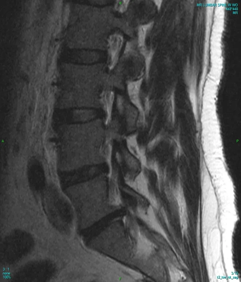
Doctor... my MRI says I have degenerative disc disease, arthritis, three bulging discs, spinal stenosis, and bone spurs. Is my back falling apart?
If you've ever asked yourself that question, you're not alone.
In fact, it's one of the most common conversations I have with patients every single week.
Many people come to my office convinced their MRI explains everything they're feeling—or worse, that the wording in the report means they're headed for surgery or permanent disability.
Fortunately, that usually isn't true.
One of the most rewarding parts of my job is watching the relief on a patient's face after we review their MRI together. Once they understand what the report actually means—and what it doesn't—the fear often begins to disappear.
That's why I wrote this guide.
My goal isn't simply to explain MRI terminology. My goal is to help you understand how spine specialists actually interpret MRI findings, why many abnormalities are a normal part of aging, and how we determine which findings are truly responsible for your pain.
By the end of this guide, I hope you'll look at your MRI with confidence instead of fear.
Why MRI Reports Sound So Scary
MRI reports are written for medical professionals.
Radiologists have one primary responsibility: describe every abnormality they see.
That means they document everything—from a tiny disc bulge to severe spinal stenosis.
They're not deciding which finding is causing your pain. They're simply describing the anatomy.
This is an important distinction.
Imagine taking your car to a mechanic for an inspection. The mechanic might notice worn tires, faded paint, a small windshield chip, and brake pads that have begun to wear.
Every one of those observations may be accurate. But if you came in because your battery died, none of those other findings explain why your car wouldn't start.
The same principle applies to your spine.
Your MRI may identify several abnormalities, but only one—or sometimes none—may actually be responsible for your symptoms.
The Biggest Misconception About Spine MRI
If there is one message I hope every patient remembers from this guide, it's this:
An abnormal MRI does not automatically mean an abnormal spine.
Modern MRI scanners are incredibly powerful.
They can detect tiny changes inside your spine long before those changes ever cause symptoms.
That's wonderful when we're looking for serious problems. But it also means MRI frequently identifies findings that are simply part of normal aging.
Over the last several decades, researchers have performed MRI scans on thousands of healthy adults who had absolutely no neck pain or back pain.
The results surprised many people.
Researchers found that a large percentage of completely pain-free adults had:
- Degenerative disc disease
- Bulging discs
- Disc dehydration
- Arthritis
- Bone spurs
- Mild spinal stenosis
In other words, many people have “abnormal” MRI findings without any pain whatsoever.
That doesn't mean your pain isn't real. It means the MRI alone rarely tells the entire story.
From Dr. Partridge
One of the biggest misconceptions I hear is: “My MRI looks terrible, so I must need surgery.” In reality, I've treated marathon runners with MRIs that looked terrible and patients with severe sciatica whose MRI showed only a tiny disc herniation. That's why I spend just as much time listening to your story and examining you as I do reviewing your MRI. The images matter. Your symptoms matter just as much.
Think of Your MRI Like Gray Hair
One of the easiest ways to understand MRI findings is to compare them to something everyone experiences: aging.
Gray hair isn't a disease. Wrinkles aren't a disease. Reading glasses aren't a disease. They're simply signs that our bodies change over time.
Your spine ages the same way.
As the years pass:
- Discs slowly lose water.
- Joints gradually develop arthritis.
- Small bone spurs form.
- Ligaments become thicker.
- The spaces around nerves may narrow.
These changes are expected. In fact, they occur in nearly everyone.
The challenge isn't identifying these changes. The challenge is determining which ones—if any—are actually causing your symptoms.
That is where experience matters.
Why Two People Can Have the Same MRI but Feel Completely Different
One of the most fascinating things about spine medicine is that two people can have nearly identical MRI scans while living completely different lives.
I've cared for patients in their 60s whose MRI reports describe severe arthritis, multiple bulging discs, and significant degeneration.
They're hiking in the mountains, golfing on weekends, and keeping up with their grandchildren.
Then I'll meet a 30-year-old with a very small disc herniation pressing on exactly the wrong nerve.
That patient may be unable to sit through dinner or sleep through the night because of severe sciatica.
Same technology. Very different stories.
That's because pain isn't determined by how impressive an MRI looks.
Pain depends on whether a particular structure is inflamed, unstable, or compressing a sensitive nerve.
That's why your MRI must always be interpreted alongside your symptoms and physical examination.
What the Research Shows
One of the most influential studies in spine medicine examined MRI scans from more than 3,000 adults with no back pain at all.
Researchers found that spinal changes become increasingly common as we age—even in people who feel completely healthy.
- Age 20: Disc degeneration is common; disc bulges are common; facet arthritis is rare.
- Age 30: Disc degeneration is more common; disc bulges are common; mild facet arthritis may be present.
- Age 40: Disc degeneration is very common; disc bulges are increasingly common; facet arthritis is common.
- Age 50: Disc degeneration is extremely common; disc bulges are very common; facet arthritis is very common.
- Age 60: Disc degeneration is nearly universal; disc bulges are very common; facet arthritis is extremely common.
- Age 70 and older: Disc degeneration is almost universal; disc bulges are extremely common; facet arthritis is nearly universal.
The takeaway is reassuring.
If you're in your 50s or 60s and your MRI mentions arthritis or degenerative changes, that doesn't necessarily mean your spine is “worn out.”
It often means your spine has simply aged—just like every other part of your body.
What This Guide Will Teach You
Over the next several sections, I'm going to walk you through your MRI the same way I explain it to patients in my office.
We'll cover:
- What every common MRI finding actually means.
- Which findings usually cause pain—and which often don't.
- The difference between a bulging disc and a herniated disc.
- Why degenerative disc disease isn't as frightening as it sounds.
- How spine specialists determine the true source of pain.
- Which MRI findings deserve urgent attention.
- Modern, evidence-based treatment options.
- When surgery may be appropriate—and when it usually isn't.
Most importantly, you'll learn how to look beyond the words on your MRI report and understand what they actually mean for you.
Our Philosophy at Horizon Spine & Pain
At Horizon Spine & Pain, we believe every patient deserves more than a copy of an MRI report.
You deserve an explanation. You deserve to understand why you're hurting. You deserve a treatment plan that's based on your symptoms, your goals, your lifestyle, and your examination—not simply a list of MRI findings.
Every week I meet patients from Payson, Spanish Fork, Springville, Mapleton, Salem, Santaquin, Provo, Orem, Lehi, and throughout Utah County who have spent months worrying about words they read on an MRI report.
My hope is that this guide changes that.
Because once you understand what your MRI is really telling you—and what it isn't—you can stop fearing the report and start focusing on what matters most:
- Finding the right diagnosis.
- Choosing the right treatment.
- Getting back to living your life.
What Does My MRI Mean?
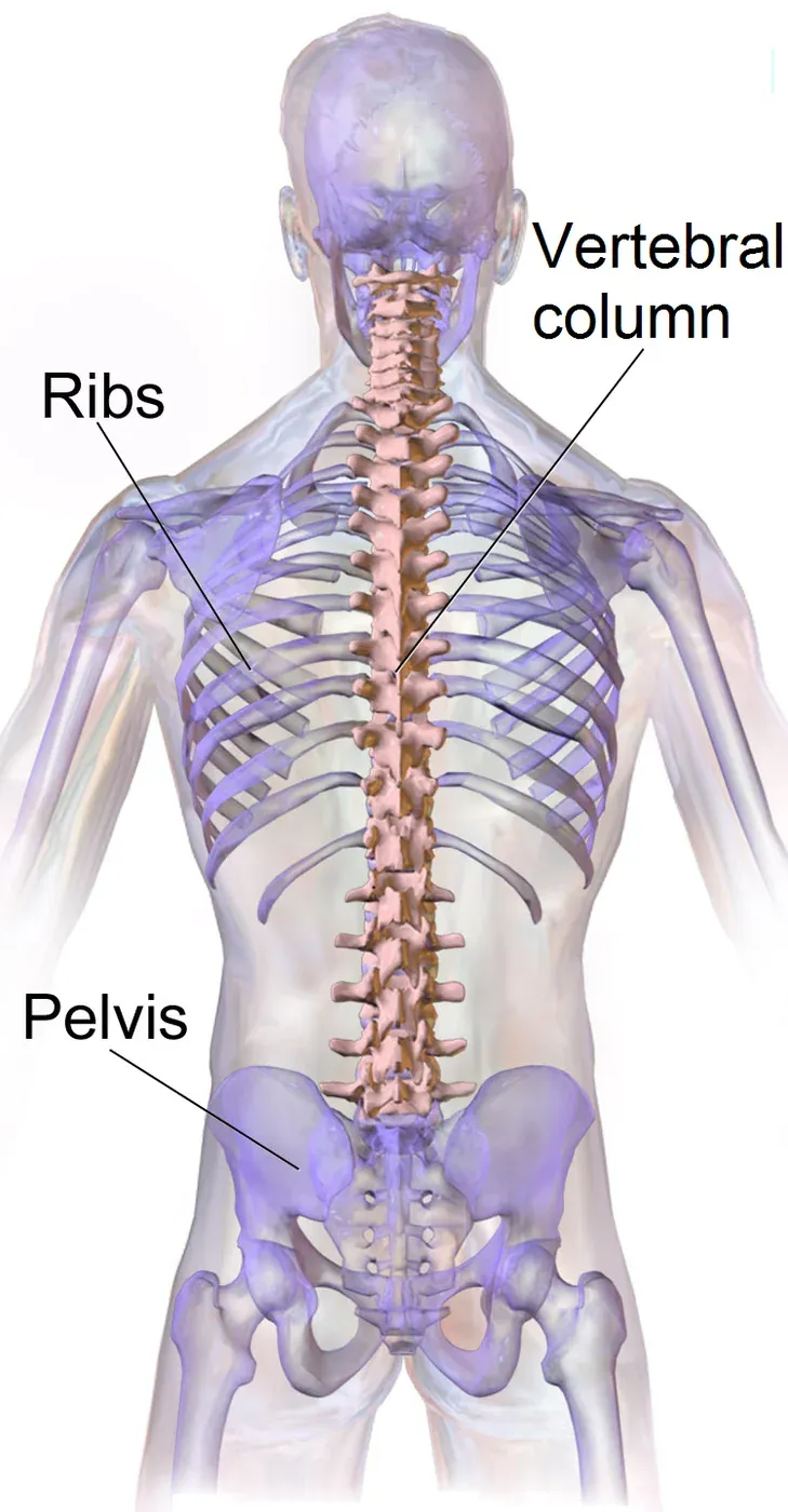
Understanding Degenerative Disc Disease, Bulging Discs, Herniated Discs, and Annular Tears
My MRI says I have degenerative disc disease. Should I be worried?
If I had to choose one diagnosis that creates more unnecessary anxiety than any other, it would probably be degenerative disc disease.
Every week I meet patients who have searched that phrase online before coming to see me. Many assume it means their spine is rapidly deteriorating or that surgery is inevitable.
Fortunately, that's almost never what it means.
Let's translate some of the most common MRI findings into plain English.
My MRI Says I Have Degenerative Disc Disease
The first thing I usually tell patients is this:
Despite the name, degenerative disc disease is usually not a disease at all.
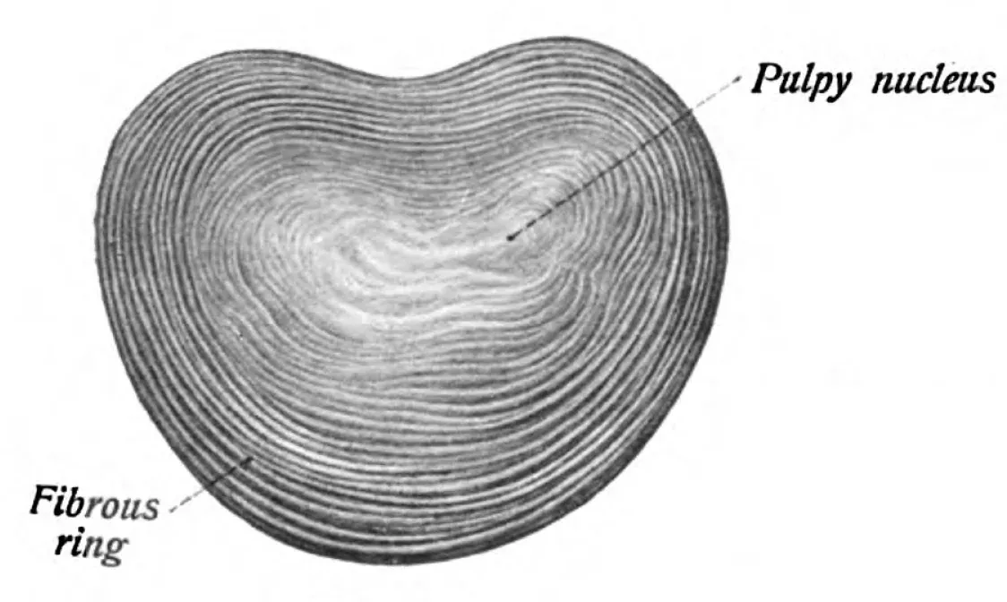
It's the medical term we use to describe the normal aging process of the discs between the bones of your spine.
Think about your hair. Over time it turns gray. Your skin develops wrinkles. Your eyesight slowly changes.
None of those things mean you're sick. They simply mean you're getting older.
Your spinal discs go through a very similar process.
When we're young, they're full of water and act like soft cushions between the vertebrae. Every time you walk, run, jump, or lift something, those discs absorb tremendous forces.
Over the years, they slowly lose some of that water content. They become a little less flexible and sometimes a little thinner. These changes happen gradually and naturally.
That's what your MRI is describing.
What Does Degenerative Disc Disease Feel Like?
Here's where things become interesting.
Many people with degenerative discs feel absolutely nothing.
Others develop:
- Aching low back pain
- Neck stiffness
- Pain after prolonged sitting
- Pain with bending forward
- Difficulty sitting for long periods
Typically, the discomfort stays in the neck or back rather than traveling down the arms or legs.
If pain begins shooting into an arm or leg, another structure—such as a herniated disc or narrowing around a nerve—is often involved.
From Dr. Partridge
One of the biggest misconceptions I hear is: “My MRI says degenerative disc disease, so my spine must be wearing out.” That's rarely how I think about it. Instead, I ask a different question: Does this particular disc actually explain your symptoms? I've seen patients with severe degeneration who are hiking every weekend. I've also treated patients whose MRI showed only mild degeneration but whose symptoms clearly pointed to a painful disc. The MRI gives us clues. Your symptoms tell us whether those clues actually matter.
What the Research Shows
One of the largest MRI studies ever performed found that disc degeneration becomes increasingly common with age—even in people who have no back pain at all.
By age 50, the majority of healthy adults have some degree of disc degeneration.
By age 60, it becomes the rule rather than the exception.
That doesn't mean everyone has back pain. It means MRI findings must always be interpreted in the context of the patient sitting in front of us.
My MRI Says Disc Desiccation
Disc desiccation sounds intimidating, but it simply means the disc has lost some of its normal water content.
Healthy discs contain a large amount of water. That's one reason they're so good at absorbing shock.
As we age, that water slowly decreases.
On MRI, healthy discs appear bright. As they lose water, they become darker.
Radiologists often describe this as:
- Disc desiccation
- Loss of hydration
- Loss of T2 signal
These all describe the same basic process.
Most of the time, disc desiccation is simply one of the earliest signs of normal aging.
Does Disc Desiccation Cause Pain?
Usually not.
Some patients with severely dehydrated discs develop chronic disc-related pain. Many others never know it's there.
The MRI finding alone doesn't tell us whether the disc is painful. That's where your symptoms become so important.
What's the Difference Between a Bulging Disc and a Herniated Disc?
This is one of the most common questions I answer in the office.
Although the two terms are often used interchangeably, they are not the same thing.
Understanding the difference can make reading your MRI much less confusing.
Bulging Disc
Imagine squeezing a jelly donut. As you apply pressure, the donut becomes slightly wider. The jelly stays inside.
That's very similar to what happens with a bulging disc.
The outer wall remains intact, but the disc extends slightly beyond its normal boundaries.
Bulging discs are incredibly common. In fact, many people have them without ever experiencing pain.
Can a Bulging Disc Cause Sciatica?
Sometimes.
If the bulging disc narrows the space where a nerve exits the spine, it may contribute to:
- Sciatica
- Arm pain
- Tingling
- Numbness
- Weakness
However, many bulging discs never touch a nerve and never require treatment.
From Dr. Partridge
One of the biggest mistakes I see is patients focusing on the words “bulging disc.” The more important question is: Where is it? A small bulge that isn't touching a nerve often causes no symptoms at all. A much smaller bulge in exactly the wrong location may produce significant pain. Location matters far more than size.
Herniated Disc
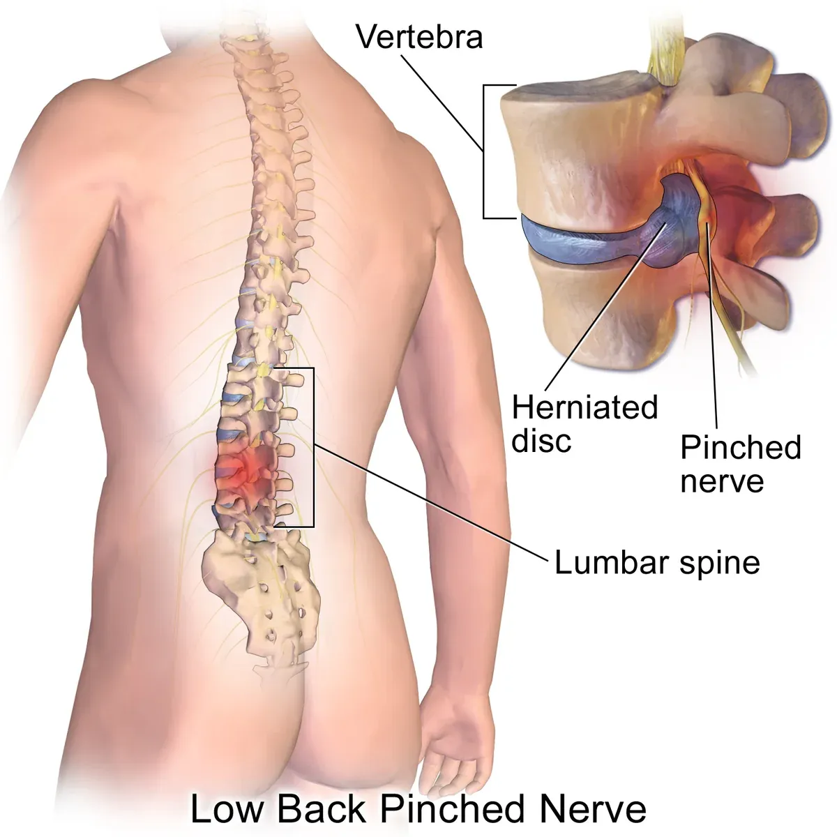
A herniated disc is different.
Instead of the entire disc bulging outward, a tear develops in the outer fibers of the disc, allowing some of the inner gel-like material to push through.
Because the disc material is now closer to nearby nerves, herniations are much more likely to produce symptoms.
Types of Disc Herniation
Disc Protrusion
The disc pushes outward, but the outer fibers still contain most of the material.
Think of it as the earliest stage of a herniation.
Disc Extrusion
The tear becomes larger, allowing disc material to extend farther beyond the disc space.
Interestingly, these larger herniations often shrink naturally over time.
Your immune system recognizes the displaced disc material as something that doesn't belong and gradually breaks it down.
Disc Sequestration
Occasionally, a fragment of disc completely separates from the parent disc.
Although this sounds frightening, many sequestrated fragments also shrink over time.
The MRI appearance may look dramatic, but surgery isn't automatically required.
What Does a Herniated Disc Feel Like?
Unlike degenerative disc disease, herniated discs often irritate nearby nerves.
Symptoms may include:
- Sharp, shooting leg pain
- Sciatica
- Burning pain
- Tingling
- Numbness
- Muscle weakness
- Pain that worsens with coughing or sneezing
Many patients say the leg pain is much worse than the back pain.
That's an important clue.
Do All Herniated Discs Need Surgery?
Absolutely not.
In fact, most don't.
Many improve significantly with:
- Time
- Physical therapy
- Anti-inflammatory medications
- Image-guided epidural steroid injections
- Activity modification
Surgery is usually reserved for patients with progressive weakness, severe neurologic deficits, bowel or bladder dysfunction, or persistent symptoms that fail to improve despite appropriate conservative treatment.
My MRI Says I Have an Annular Tear
Despite the name, an annular tear doesn't necessarily mean your disc has ripped apart.
The annulus is the tough outer layer of the disc.
Over time—or sometimes after an injury—small cracks can develop within its fibers.
Radiologists may describe these as:
- Annular fissures
- Annular tears
- High-intensity zones (HIZ)
These small tears are actually fairly common.
Some become painful because the outer portion of the disc contains pain-sensitive nerve fibers. Others are completely painless.
This is another example of why MRI findings must always be interpreted alongside your symptoms.
From Dr. Partridge
One of the questions patients ask me most is: “If I have a tear in my disc, does that mean it's going to rupture?” Fortunately, that's usually not the case. Most annular fissures remain stable over time. Some heal. Some become asymptomatic. Others contribute to chronic disc pain. The MRI tells me the tear exists. Your history and examination tell me whether it's actually important.
Arthritis, Spinal Stenosis, and Other Common MRI Findings Explained
Understanding the MRI Terms That Confuse Patients the Most
By the time many patients get to this point in their MRI report, they've already read words like degenerative disc disease, bulging disc, or herniated disc.
Then the report continues:
- Facet arthropathy
- Ligamentum flavum hypertrophy
- Foraminal stenosis
- Spondylolisthesis
For most people, that's when everything starts sounding like another language.
The good news? These terms are much easier to understand than they sound.
Let's translate them into plain English.
My MRI Says I Have Arthritis in My Spine
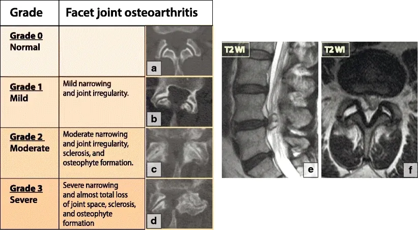
Most people are surprised to learn that the spine contains dozens of joints.
Just like your knees, hips, shoulders, and fingers, those joints can develop arthritis over time.
They're called facet joints.
Every vertebra connects to the one above and below through a pair of these small joints, allowing your spine to bend, twist, and move while maintaining stability.
Like every other joint in your body, they gradually experience wear over the years.
That's completely normal.
What Does Facet Arthritis Feel Like?
Facet arthritis usually produces a very different type of pain than a herniated disc.
Patients commonly describe:
- Aching pain across the low back
- Pain that stays close to the spine
- Morning stiffness
- Pain after standing for long periods
- Pain when leaning backward
- Difficulty twisting or looking over one shoulder
Unlike sciatica, the pain usually doesn't shoot below the knee or travel all the way into the hand.
Instead, it tends to remain in the neck, shoulders, low back, or buttocks.
From Dr. Partridge
One of the most common things I hear is: “My MRI says I have arthritis. Does that mean nothing can be done?” Absolutely not. Facet arthritis is actually one of the most treatable causes of chronic spine pain. When a patient's symptoms, examination, and MRI all point toward the facet joints, we can often confirm the diagnosis with medial branch blocks. If those blocks provide significant temporary relief, radiofrequency ablation may provide many months—and sometimes more than a year—of meaningful pain reduction. The MRI identifies the arthritis. The diagnostic block tells us whether it's actually causing the pain.
Bone Spurs (Osteophytes)
Another word that often worries patients is osteophyte.
Fortunately, osteophytes are simply bone spurs.
Bone spurs develop because the body is incredibly smart.
When a joint becomes unstable or experiences increased stress over many years, the body often responds by laying down extra bone in an attempt to stabilize that area.
It's almost like reinforcing an old bridge.
Unfortunately, that extra bone sometimes narrows the spaces around nearby nerves. When that happens, symptoms may develop.
Many bone spurs never cause problems. Others become important because of where they form—not because they exist.
My MRI Says I Have Spinal Stenosis
This is probably one of the most misunderstood diagnoses in spine care.
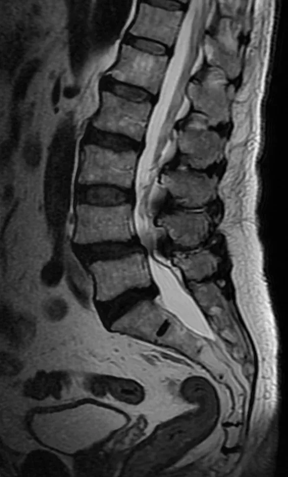
The word stenosis simply means narrowing. Nothing more.
Think of your spinal canal like a highway. Your spinal cord and spinal nerves travel through that highway.
Over time, several age-related changes may gradually reduce the available space:
- Bulging discs
- Arthritis
- Bone spurs
- Thickened ligaments
Eventually the highway becomes narrower. That's spinal stenosis.
Why Does Spinal Stenosis Hurt?
Interestingly, spinal stenosis doesn't always hurt.
Some people have severe narrowing with very few symptoms.
Others develop a very characteristic pattern known as neurogenic claudication.
Patients often tell me:
I can stand for only a few minutes before my legs start aching.
Walking through Costco is miserable unless I'm leaning over the shopping cart.
As soon as I sit down, my legs feel better.
Those are classic descriptions of spinal stenosis.
When you lean forward, the spinal canal opens slightly, giving the nerves more room.
That's why leaning on a shopping cart often provides relief.
Does Everyone With Stenosis Need Surgery?
No.
In fact, many people with mild or even moderate spinal stenosis do very well with conservative treatment.
Depending on the severity of symptoms, treatment may include:
- Physical therapy
- Activity modification
- Weight management
- Epidural steroid injections
- The MILD procedure for carefully selected patients with ligament-related narrowing
- Surgical decompression when symptoms become severe or progressive
The decision isn't based solely on the MRI. It's based on how much the narrowing affects your quality of life.
From Dr. Partridge
One MRI finding I pay close attention to isn't simply how narrow the canal looks. It's whether that narrowing explains what the patient experiences every day. I've seen MRIs with severe stenosis in patients who still hike every weekend. I've also treated patients with only moderate narrowing who could barely make it across the grocery store. The MRI gives us part of the answer. Your story completes it.
Foraminal Stenosis
If central stenosis narrows the main highway, foraminal stenosis narrows the exit ramps.
Each spinal nerve leaves the spine through a small opening called the neural foramen.
When arthritis, disc bulges, or bone spurs narrow that opening, the nerve may become compressed.
This is one of the most common causes of:
- Sciatica
- Arm pain
- Numbness
- Tingling
- Muscle weakness
Unlike central stenosis, foraminal stenosis often affects one specific nerve. That means the symptoms usually follow a predictable pattern.
For example:
- An L5 nerve root typically produces pain traveling down the outside of the leg into the top of the foot.
- An S1 nerve root often produces pain into the back of the calf and the bottom of the foot.
Those patterns help physicians match MRI findings with symptoms.
Ligamentum Flavum Hypertrophy
This is another phrase that sounds much more alarming than it really is.
The ligamentum flavum is a strong ligament running along the back of the spinal canal.
As we age, it often becomes thicker.
This thickening is extremely common. By itself, it may not cause symptoms.
However, when combined with arthritis and disc degeneration, it can contribute to spinal stenosis by taking up additional space within the spinal canal.
My MRI Says I Have Spondylolisthesis
Spondylolisthesis simply means that one vertebra has slipped slightly forward relative to the one beneath it.
Sometimes this develops because of arthritis. Sometimes it's related to a stress fracture that occurred years earlier.
Many patients never know they have it.
Others develop:
- Mechanical low back pain
- Leg pain
- Spinal stenosis
- Difficulty standing or walking
The amount of slippage doesn't always predict how much pain someone experiences.
Retrolisthesis
Retrolisthesis is simply the opposite.
Instead of slipping forward, one vertebra slips slightly backward.
Most cases are mild. Many require no specific treatment.
The finding becomes important only when it contributes to instability or nerve compression.
Synovial Cysts
Facet joints contain a small amount of lubricating fluid.
Occasionally that fluid forms a small sac called a synovial cyst.
Many synovial cysts never cause symptoms.
However, if the cyst grows large enough to compress a nearby nerve, patients may develop:
- Sciatica
- Leg numbness
- Weakness
- Difficulty walking
Treatment depends on symptoms and may include observation, injections, aspiration in selected cases, or surgery when significant nerve compression is present.
Schmorl's Nodes
Schmorl's nodes are among the most common incidental MRI findings.
They occur when a small amount of disc material pushes into the bone above or below the disc.
Most patients never know they have them. Most require no treatment.
Occasionally, a newly developed Schmorl's node may become temporarily painful, but the vast majority are simply signs of previous disc degeneration.
Vertebral Hemangiomas
Another MRI finding that frequently causes unnecessary anxiety is the vertebral hemangioma.
Despite the intimidating name, these are usually benign collections of blood vessels inside a vertebra.
They're extremely common.
Most people never develop symptoms. Most never require treatment.
Unless your physician tells you otherwise, a vertebral hemangioma is usually an incidental finding rather than the cause of pain.
Tarlov Cysts
Tarlov cysts are fluid-filled sacs that develop around nerve roots near the lower spine.
They're discovered incidentally on many MRI scans.
Most never produce symptoms. Only a small percentage become clinically significant.
Determining whether a Tarlov cyst is actually responsible for symptoms requires careful evaluation because many patients have cysts that are completely unrelated to their pain.
Bertolotti Syndrome
One MRI finding many patients—and even some healthcare providers—have never heard of is Bertolotti syndrome.
Some people are born with an enlarged transverse process on the lowest lumbar vertebra that partially connects with the sacrum.
Most never develop symptoms.
In some individuals, however, this extra connection alters spinal mechanics and becomes a source of chronic low back pain.
Because it's relatively uncommon, the diagnosis is sometimes overlooked.
Incidental MRI Findings That Usually Aren't Dangerous
MRI reports often include findings that sound intimidating but are simply part of normal anatomy or aging.
Examples include:
- Mild scoliosis
- Small hemangiomas
- Schmorl's nodes
- Mild retrolisthesis
- Mild disc bulges
- Early facet arthritis
- Small Tarlov cysts
These findings deserve to be documented, but they don't automatically explain pain.
This is another reminder that MRI reports must always be interpreted in the context of the whole patient.
From Dr. Partridge
One of the greatest values of an experienced spine specialist isn't simply recognizing abnormalities on an MRI. It's recognizing which abnormalities don't matter. Every MRI contains findings. Not every finding deserves treatment. One of my responsibilities is helping patients understand the difference so we focus on the problem that's actually limiting their life—not simply the one with the most impressive medical terminology.
Key Takeaways
If you've made it this far, you've probably realized something important:
Many MRI findings sound much worse than they actually are.
Arthritis is common. Disc degeneration is common. Bone spurs are common. Even spinal stenosis is common as we age.
The real question isn't whether those findings exist. The question is whether they explain your symptoms.
That's why experienced spine specialists combine your MRI with your physical examination, your medical history, and sometimes diagnostic injections before recommending treatment.
We don't treat MRI reports. We treat people.
Does Your MRI Actually Explain Your Pain?
How Spine Specialists Connect MRI Findings to Symptoms
By now, you've probably noticed a common theme throughout this guide.
Not every MRI finding causes pain.
In fact, one of the biggest challenges in spine medicine isn't finding abnormalities on an MRI—it's determining which abnormality is actually responsible for your symptoms.
That may sound surprising.
After all, if an MRI shows three bulging discs, arthritis, spinal stenosis, and bone spurs, shouldn't those findings explain why your back hurts?
Sometimes they do. Sometimes they don't.
This is where experience becomes incredibly important.
Reading an MRI report is only the beginning. The real job is putting together all the pieces of the puzzle.
Think of Your Diagnosis Like a Puzzle
Imagine dumping a 1,000-piece puzzle onto a table.
The MRI is one piece. Your symptoms are another. Your physical examination is another. Your medical history is another.
How your pain started matters. What makes it better matters. What makes it worse matters. Your age matters. Your activity level matters. Previous surgeries matter.
Sometimes even your response to a previous injection becomes another important piece.
Only when all of those pieces fit together can we confidently identify the true source of pain.
One missing piece can completely change the picture.
The Three Questions I Ask Every Time I Review an MRI
1. Does the MRI Match the Symptoms?
This is always my first question.
Let's imagine two patients.
Patient A
- Pain begins in the lower back.
- It shoots down the outside of the leg.
- It travels into the top of the foot.
- There's numbness in the big toe.
- The MRI shows a right L4-L5 foraminal disc herniation compressing the L5 nerve root.
Everything matches.
The patient's story and the MRI are telling the same story.
That gives me confidence we're treating the correct problem.
Now consider someone else.
Patient B
- Pain travels down the left leg.
- The MRI shows only a right-sided disc herniation.
Those findings don't fit together.
The MRI may still contain an abnormality, but it probably isn't the cause of this patient's symptoms.
Before recommending treatment, I want the MRI and the patient's story to point in the same direction.
2. Does the Physical Examination Match the MRI?

Your physical examination is just as important as your MRI.
During your visit, I evaluate:
- Muscle strength
- Sensation
- Reflexes
- Walking pattern
- Balance
- Range of motion
- Areas of tenderness
- Which movements reproduce your pain
These findings often confirm—or challenge—what we see on imaging.
For example, if an MRI shows severe nerve compression but your neurological examination is completely normal and your symptoms don't follow that nerve's pattern, I may question whether the MRI finding is truly responsible.
On the other hand, if your examination perfectly matches the MRI, we gain much more confidence in the diagnosis.
3. Could Something Else Be Causing the Pain?
One of the biggest misconceptions is that every pain problem originates in the spine.
That's simply not true.
Several conditions can mimic spinal disorders, including:
- Hip arthritis
- Sacroiliac joint dysfunction
- Shoulder injuries
- Peripheral nerve entrapment
- Knee pathology
- Vascular disease
- Muscle injuries
- Tendon disorders
One of my responsibilities is making sure we're treating the right structure—not simply the most obvious MRI finding.
From Dr. Partridge
One of the most valuable things I do during an evaluation isn't identifying what is causing pain. It's identifying what isn't. Patients are often surprised when I tell them, “I don't think this MRI finding is your problem.” Sometimes that's the most important thing I can say. Because once we stop chasing the wrong diagnosis, we can focus on finding the right one.
Why Bigger Doesn't Always Mean Worse
Patients often assume the largest abnormality on an MRI must be the most painful.
Medicine doesn't always work that way.
A massive central disc bulge may produce surprisingly few symptoms if it isn't compressing sensitive nerves.
Meanwhile, a tiny disc herniation inside the neural foramen may cause severe sciatica because it's pressing directly against a nerve root.
Location matters. Inflammation matters. Nerve sensitivity matters.
That's why size alone rarely determines treatment.
Why Pain Doesn't Always Equal Damage
Pain is much more complicated than most people realize.
Think about getting a paper cut. The injury is tiny. Yet it can hurt tremendously.
Now think about someone with advanced arthritis who experiences very little discomfort.
Pain depends on much more than structural damage.
Factors that influence pain include:
- Inflammation
- Nerve irritation
- Sleep quality
- Physical conditioning
- Previous injuries
- Emotional stress
- Central nervous system sensitivity
This is one reason why two people with identical MRIs can experience completely different levels of pain.
Can You Have Severe Pain With a Normal MRI?
Absolutely.
Some painful conditions produce very little change on MRI.
Examples include:
- Sacroiliac joint dysfunction
- Certain muscular injuries
- Myofascial pain
- Early disc injuries
- Some ligament injuries
- Peripheral nerve disorders
MRI is one of the best diagnostic tools available. But no imaging study is perfect.
That's why physicians never diagnose pain based solely on pictures.
Can You Have a Terrible MRI and Feel Great?
Absolutely.
In fact, I see this regularly.
Many active adults have MRIs showing:
- Multiple bulging discs
- Severe arthritis
- Disc degeneration
- Mild spinal stenosis
Yet they continue to golf, ski, hike, cycle, and live active lives with very little pain.
This is one of the reasons I encourage patients not to panic after reading their MRI report.
The wording often sounds much worse than the reality.
Understanding Nerve Pain
One of the most useful clues in spine medicine is the pattern of pain.
Different nerves produce pain in different locations.
For example:
Cervical Spine
- C5: Pain in the shoulder; weakness lifting the arm.
- C6: Pain into the thumb; possible weakness bending the elbow.
- C7: Pain into the middle finger; difficulty straightening the elbow.
- C8: Pain into the ring and little fingers; weakness with grip strength.
Lumbar Spine
- L3: Pain into the front of the thigh; difficulty climbing stairs.
- L4: Pain around the knee and inner leg; weakness straightening the knee; reduced patellar reflex.
- L5: Pain down the outside of the leg and into the top of the foot; difficulty lifting the foot or big toe.
- S1: Pain down the back of the leg and into the bottom of the foot; difficulty standing on the toes; reduced Achilles reflex.
These predictable patterns help us determine whether MRI findings actually match the patient's symptoms.
When Diagnostic Injections Become Helpful
Sometimes the MRI and examination still leave questions unanswered.
This is where diagnostic injections can be extremely valuable.
Unlike therapeutic injections, which are designed primarily to reduce pain, diagnostic injections help identify the true pain generator.
Examples include:
Medial Branch Blocks
Used to determine whether facet joints are causing pain.
Selective Nerve Root Blocks
Used when multiple nerves appear abnormal on MRI and we need to identify which one is responsible for symptoms.
Sacroiliac Joint Injections
Helpful when SI joint pain closely mimics lumbar spine disorders.
If pain improves dramatically after numbing a specific structure, it provides strong evidence that we've identified the correct diagnosis.
From Dr. Partridge
One of my favorite sayings is: “The best treatment starts with the right diagnosis.” Sometimes the most valuable procedure I perform isn't designed to treat pain. It's designed to answer one question: “Where is this pain actually coming from?” Once we know that answer, treatment becomes much more precise.
MRI Red Flags: When You Should Seek Immediate Medical Attention
Most MRI findings are not emergencies.
However, certain symptoms deserve immediate evaluation.
Seek prompt medical attention if you experience:
- Loss of bowel or bladder control.
- Numbness in the groin or saddle area.
- Rapidly worsening weakness.
- Difficulty walking because of new neurological problems.
- Fever with severe back pain.
- Significant trauma followed by severe pain.
- Unexplained weight loss with persistent back pain.
- A history of cancer with new severe spine pain.
Fortunately, these situations are uncommon, but recognizing them is extremely important.
The Bottom Line
One of the most important lessons in spine medicine is that MRI findings don't exist in isolation.
They must fit the patient's symptoms. They must fit the physical examination. They must fit the neurological findings.
Only then can we confidently identify the true pain generator.
At Horizon Spine & Pain, that's exactly how we approach every patient.
We don't simply review MRI reports. We take the time to understand your story.
Because the goal isn't to explain your MRI. The goal is to explain your pain.
What's Next? Modern Treatment Options, Common Questions, and My Final Thoughts
By now, you've learned something that surprises many people:
An MRI report is not a treatment plan.
It's one piece of information. A very important piece—but only one piece.
The goal of modern spine care isn't to “fix” every abnormal MRI finding. The goal is to identify the structure that's actually causing your symptoms and choose the least invasive treatment that helps you return to the life you enjoy.
Fortunately, that's exactly where we have made tremendous progress over the last two decades.
Many patients who once would have been told they needed surgery now have excellent nonsurgical options available.
Let's talk about what those treatments are—and when they make sense.
Treatment Should Match the Diagnosis
One of the biggest mistakes in spine care is recommending treatment based solely on an MRI report.
Instead, treatment should always answer three questions:
- What structure is causing the pain?
- How much is it affecting your quality of life?
- Which treatment has the best evidence for this specific diagnosis?
Those questions guide every recommendation I make.
Physical Therapy: The Foundation of Spine Care

Many people are disappointed when they hear a physician recommend physical therapy.
They were expecting an injection or a procedure.
The reality is that physical therapy is one of the most effective long-term treatments for many spine conditions.
A good therapy program can help:
- Improve flexibility
- Restore normal movement
- Strengthen the muscles supporting the spine
- Improve posture
- Increase endurance
- Reduce future injuries
- Build confidence with movement
The goal isn't simply to reduce pain today.
The goal is to improve how your spine functions for years to come.
From Dr. Partridge
One of the biggest myths I hear is: “If it hurts, I should stop moving.” While there are certainly exceptions, prolonged inactivity often makes chronic spine pain worse. The right kind of movement is frequently one of the best treatments we have.
Lifestyle Changes Matter More Than Most People Realize
Although there isn't one perfect diet or exercise program for spine pain, there are several habits that consistently improve outcomes.
These include:
- Maintaining a healthy body weight
- Avoiding nicotine products
- Walking regularly
- Strength training when appropriate
- Prioritizing quality sleep
- Managing stress
- Staying physically active
These recommendations may sound simple, but they are supported by decades of research.
Healthy muscles support healthy joints. Healthy joints protect healthy nerves.
Medications: Helpful Tools, Not Complete Solutions
Medications can reduce pain and inflammation, allowing patients to stay active while healing.
Depending on the diagnosis, treatment may include:
- Acetaminophen
- Nonsteroidal anti-inflammatory medications (NSAIDs)
- Neuropathic pain medications for nerve-related pain
- Muscle relaxants for selected patients
- Topical medications
- Short courses of oral steroids in carefully selected situations
Medication decisions should always balance potential benefits with possible risks and side effects.
For many chronic spine conditions, medications work best when combined with exercise, education, and targeted rehabilitation.
Image-Guided Spine Procedures
When conservative treatments aren't enough, minimally invasive procedures may help reduce pain while allowing patients to continue working, exercising, and participating in daily life.
These procedures are most effective when they are directed at the correct pain generator.
Epidural Steroid Injections
Best suited for:
- Lumbar radiculopathy
- Cervical radiculopathy
- Disc herniations
- Sciatica
- Selected patients with spinal stenosis
The goal is to reduce inflammation around an irritated nerve.
These injections do not “fix” the disc itself, but they often decrease nerve irritation enough to allow the body to heal while patients continue rehabilitation.
Medial Branch Blocks and Radiofrequency Ablation
Facet arthritis is one of the most common causes of chronic neck and back pain.
When the history, examination, and MRI suggest facet-mediated pain, diagnostic medial branch blocks help confirm the diagnosis.
If those blocks provide significant temporary relief, radiofrequency ablation (RFA) may offer longer-lasting improvement by interrupting pain signals from the small nerves supplying the facet joints.
Many patients experience six to eighteen months of relief before the nerves regenerate.
From Dr. Partridge
One of the reasons I like diagnostic blocks is that they help answer an important question before we perform a longer-lasting treatment: “Are we treating the correct structure?” When the diagnosis is right, outcomes are almost always better.
Sacroiliac Joint Injections
Not every patient with low back pain has a lumbar spine problem.
The sacroiliac (SI) joint is a common—and often overlooked—source of pain.
Symptoms frequently include:
- Pain below the beltline
- Pain over one buttock
- Difficulty standing from a chair
- Pain climbing stairs
- Pain rolling over in bed
Image-guided SI joint injections help both diagnose and treat this condition.
Patients with persistent SI joint pain may also benefit from minimally invasive SI joint stabilization after appropriate evaluation.
Basivertebral Nerve Ablation (Intracept)
Some patients develop chronic vertebrogenic low back pain associated with Modic type I or II changes on MRI.
For carefully selected individuals, basivertebral nerve ablation (Intracept) targets the nerve supplying the vertebral endplates rather than the disc itself.
Clinical trials have demonstrated meaningful improvements in pain and function for appropriately selected patients.
Kyphoplasty
Vertebral compression fractures often cause sudden, severe back pain, especially in patients with osteoporosis.
When appropriate, kyphoplasty can stabilize the fracture, reduce pain, and help patients return to normal activities more quickly.
Spinal Cord Stimulation
For some patients with chronic neuropathic pain—particularly after previous spine surgery or in conditions such as complex regional pain syndromes—spinal cord stimulation may reduce pain and improve quality of life.
Modern systems are smaller, more sophisticated, and more customizable than ever before.
A temporary trial is performed before permanent implantation to ensure the therapy is effective.
When Is Surgery the Right Choice?
One of the biggest misconceptions I encounter is that surgery is either the first option or the last option.
Neither is true.
Surgery is simply one treatment among many.
It may be the best option when:
- Progressive weakness develops
- Significant nerve compression persists despite conservative care
- Severe spinal stenosis limits walking
- Spinal instability is present
- Certain fractures require stabilization
- Tumors or infections require surgical treatment
- Bowel or bladder dysfunction suggests severe nerve compression
For many other conditions, nonsurgical treatment is highly successful.
The decision should always be individualized.
Frequently Asked Questions
If My MRI Looks Bad, Does That Mean I Need Surgery?
No.
Many patients with severe MRI findings improve with nonsurgical treatment.
Your symptoms and examination are far more important than the wording of the MRI report alone.
Can a Herniated Disc Heal Without Surgery?
Yes.
Many herniated discs shrink naturally over time as the body gradually breaks down and absorbs the displaced disc material.
Is It Safe to Exercise if I Have Arthritis in My Spine?
For most people, yes.
Appropriately prescribed exercise is one of the most effective treatments for chronic spine pain.
Remaining active is usually far better than prolonged rest.
Why Does My MRI Sound So Much Worse Than I Feel?
MRI scanners are incredibly sensitive.
They frequently identify age-related changes that have little or nothing to do with symptoms.
That's why MRI findings must always be interpreted alongside your history and physical examination.
Can I Have Severe Pain With Only a “Mild” MRI?
Absolutely.
A small disc herniation pressing directly on a nerve may cause severe pain.
Meanwhile, another patient with extensive arthritis may have very few symptoms.
Pain depends on much more than how dramatic the MRI appears.
My Final Thoughts

If you've made it this far, I hope you've come away with one important message:
Your MRI is part of your story—but it isn't the whole story.
As physicians, it's easy to focus on images.
As patients, it's easy to focus on words like degeneration, stenosis, or arthritis.
But neither tells the complete story by itself.
Every day, I meet patients throughout Utah County who have spent months worrying about an MRI report that they didn't fully understand.
Many are relieved to discover that the findings they feared most are actually common signs of aging.
Others finally receive an explanation for symptoms they've been living with for years.
Both conversations are equally rewarding.
My goal has never been to treat MRI reports.
My goal is to understand the person sitting in front of me.
That means listening carefully, performing a thoughtful examination, reviewing imaging in context, and building a treatment plan that fits your goals—not just your scan.
Whether your treatment involves exercise, physical therapy, an image-guided procedure, surgery, or simply reassurance, it should always begin with an accurate diagnosis.
I hope this guide has helped you better understand your MRI, reduced some of the uncertainty surrounding your report, and given you confidence that effective, evidence-based treatment options are available.
Thank you for taking the time to read it.
I wish you the very best on your journey toward recovery.
Pain relief is on the horizon.
—Dr. Devan Partridge
Key References
The recommendations and educational information in this guide are based on current evidence from peer-reviewed research and national specialty organizations, including:
- Brinjikji W, et al. Systematic Literature Review of Imaging Features of Spinal Degeneration in Asymptomatic Populations. American Journal of Neuroradiology. 2015.
- Jensen MC, et al. Magnetic Resonance Imaging of the Lumbar Spine in People Without Back Pain. New England Journal of Medicine. 1994.
- Boden SD, et al. Abnormal Magnetic Resonance Scans of the Lumbar Spine in Asymptomatic Subjects. Journal of Bone and Joint Surgery. 1990.
- Foster NE, et al. Prevention and Treatment of Low Back Pain: Evidence, Challenges, and Promising Directions. The Lancet. 2018.
- Hartvigsen J, et al. What Low Back Pain Is and Why We Need to Pay Attention. The Lancet. 2018.
- Chou R, et al. Interventions for Acute and Chronic Low Back Pain. Journal of Orthopaedic & Sports Physical Therapy. 2021.
- North American Spine Society (NASS). Evidence-Based Clinical Guidelines.
- American College of Radiology (ACR). Appropriateness Criteria for Low Back Pain.
Medical Disclaimer
This guide is intended for educational purposes only and should not replace an evaluation by a qualified healthcare professional. Every patient's condition is unique, and treatment recommendations should always be individualized based on your history, examination, imaging findings, and overall health.